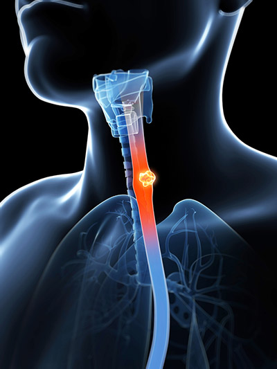
Oesophageal Cancer
How common is oesophageal cancer?
 The incidence of oesophageal cancer has been steadily rising over the last 10 years. It usually affects the 50-70 year age group and is more common in males than females. In the UK, the majority of oesophageal cancers are adenocarcinomas, which arise around the join between the oesophagus and stomach. The other main type is squamous cell carcinoma, which usually occurs higher up in the oesophagus.
Who does it effect?
The rise in incidence of oesophageal adenocarcinoma, is thought to be related to gastro-oesophageal reflux disease. Squamous cell cancers are caused by smoking and dietary factors.
How is it detected?
The majority of patients will present with difficulty in swallowing. Any patients with swallowing difficulties or new reflux symptoms should be urgently investigated.
Some cancers are detected whilst patients are under surveillance for a condition known as Barrett’s Oesophagus. This is a change in the lining of the oesophagus due to the high exposure of the lower oesophagus to gastro-oesophageal reflux. Patients with this condition are more likely to develop oesophageal cancer and are usually regularly checked by endoscopy.
How is the diagnosis made?
The first investigation for swallowing difficulties is an endoscopy. Biopsies are taken and sent to the laboratory for analysis. It usually takes 2-3 days to get a result. Once the diagnosis has been made a series of investigations are performed to gain an understanding of the extent of the cancer (cancer staging).
- CT Scan – A CT scan is a quick painless procedure to obtain pictures of the body. Contrast will be given by injection during the procedure to highlight the blood vessels. It usually takes about 15mins.
- PET scan – This is a similar procedure to the CT scan and can sometimes be combined. Sugar is labeled and then administered via injection. Areas of high activity in the body take up the sugar and are then detected by the scanner. It is used to detect any focus of cancer not picked up on the CT scan.
- EUS – This is a similar procedure to the endoscopy, except the probe has an ultrasound device on the end, which enables the doctor to examine the cancer and surrounding tissue in precise detail. It takes longer than an endoscopy and may require more sedation.
- Staging Laparoscopy – If the cancer is near the stomach then a keyhole inspection of the abdomen is performed. Occasionally scans do not detect small foci of cancer cells, which are easily seen when viewed with a keyhole camera. The procedure is quick and patients go home a few hours after the procedure.
How is the oesophageal cancer treated?
The staging tests outlined above enable us to understand how far the cancer has spread. If the cancer remains confined to the oesophagus then a curative approach is used. Cancer, which has spread away from the oesophagus to other organs, may not be curable and will require chemotherapy to attempt to gain control of the disease.
If the cancer is very early, it can be treated by endoscopy, but this is uncommon. The majority of curable cancers will require a combination of chemotherapy and surgery to obtain the best results. In certain situations radiotherapy will also be considered for further treatment.


